Back Raphe-Kerne German Núcleos del rafe Spanish هسته رافه Persian Raphe-tumake Finnish Noyaux du raphé French 縫線核 Japanese 솔기핵 Korean Nuclei raphes Latin Nuclei raphes Dutch Jądra szwu Polish
| Raphe nuclei | |
|---|---|
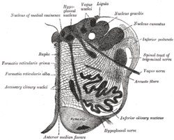
 Section of the medulla oblongata at about the middle of the olive. (Raphe nuclei not labeled, but 'raphe' labeled at left.) | |
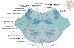
 Horizontal cross section of the brainstem at the lower pons. The raphe nucleus is labeled #18 in the middle. | |
| Details | |
| Identifiers | |
| Latin | nuclei raphe |
| MeSH | D011903 |
| NeuroLex ID | nlx_anat_20090205 |
| TA98 | A14.1.04.257 A14.1.04.318 A14.1.05.402 A14.1.05.601 A14.1.06.401 |
| TA2 | 6035, 5955 |
| FMA | 84017 |
| Anatomical terms of neuroanatomy | |
The raphe nuclei (Greek: ῥαφή, "seam")[1] are a moderate-size cluster of nuclei found in the brain stem. They have 5-HT1 receptors which are coupled with Gi/Go-protein-inhibiting adenyl cyclase. They function as autoreceptors in the brain and decrease the release of serotonin. The anxiolytic drug Buspirone acts as partial agonist against these receptors.[2] Selective serotonin reuptake inhibitor (SSRI) antidepressants are believed to act in these nuclei, as well as at their targets.[3]
- ^ Liddell HG, Scott R (1940). A Greek-English Lexicon. Oxford: Clarendon Press.
revised and augmented throughout by Sir Henry Stuart Jones with the assistance of Roderick McKenzie
- ^ Siegel GJ, Agranoff BW, Fisher SK, Albers RW, Uhler MD (1999). "Understanding the neuroanatomical organization of serotonergic cells in brain provides insight into the functions of this neurotransmitter". Basic Neurochemistry (Sixth ed.). Lippincott Williams and Wilkins. ISBN 978-0-397-51820-3.
In 1964, Dahlstrom and Fuxe (discussed in [2]), using the Falck-Hillarp technique of histofluorescence, observed that the majority of serotonergic soma are found in cell body groups, which previously had been designated as the raphe nuclei.
- ^ Briley M, Moret C (October 1993). "Neurobiological mechanisms involved in antidepressant therapies". Clinical Neuropharmacology. 16 (5): 387–400. doi:10.1097/00002826-199310000-00002. PMID 8221701.