Back هيكل خلوي Arabic Цитоскелет Bulgarian কোষকঙ্কাল Bengali/Bangla Citoskelet BS Citoesquelet Catalan پەیکەری خانە CKB Cytoskelet Czech Cytosgerbwd Welsh Cytoskelet Danish Cytoskelett German
| Cell biology | |
|---|---|
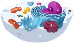
| Animal cell diagram | |
 Components of a typical animal cell:
|

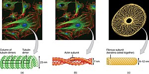
The cytoskeleton is a complex, dynamic network of interlinking protein filaments present in the cytoplasm of all cells, including those of bacteria and archaea.[2] In eukaryotes, it extends from the cell nucleus to the cell membrane and is composed of similar proteins in the various organisms. It is composed of three main components: microfilaments, intermediate filaments, and microtubules, and these are all capable of rapid growth or disassembly depending on the cell's requirements.[3]
A multitude of functions can be performed by the cytoskeleton. Its primary function is to give the cell its shape and mechanical resistance to deformation, and through association with extracellular connective tissue and other cells it stabilizes entire tissues.[4][5] The cytoskeleton can also contract, thereby deforming the cell and the cell's environment and allowing cells to migrate.[6] Moreover, it is involved in many cell signaling pathways and in the uptake of extracellular material (endocytosis),[7] the segregation of chromosomes during cellular division,[4] the cytokinesis stage of cell division,[8] as scaffolding to organize the contents of the cell in space[6] and in intracellular transport (for example, the movement of vesicles and organelles within the cell)[4] and can be a template for the construction of a cell wall.[4] Furthermore, it can form specialized structures, such as flagella, cilia, lamellipodia and podosomes. The structure, function and dynamic behavior of the cytoskeleton can be very different, depending on organism and cell type.[4][9][8] Even within one cell, the cytoskeleton can change through association with other proteins and the previous history of the network.[6]
A large-scale example of an action performed by the cytoskeleton is muscle contraction. This is carried out by groups of highly specialized cells working together. A main component in the cytoskeleton that helps show the true function of this muscle contraction is the microfilament. Microfilaments are composed of the most abundant cellular protein known as actin.[10] During contraction of a muscle, within each muscle cell, myosin molecular motors collectively exert forces on parallel actin filaments. Muscle contraction starts from nerve impulses which then causes increased amounts of calcium to be released from the sarcoplasmic reticulum. Increases in calcium in the cytosol allows muscle contraction to begin with the help of two proteins, tropomyosin and troponin.[10] Tropomyosin inhibits the interaction between actin and myosin, while troponin senses the increase in calcium and releases the inhibition.[11] This action contracts the muscle cell, and through the synchronous process in many muscle cells, the entire muscle.
- ^
 This article incorporates text available under the CC BY 4.0 license. Betts, J Gordon; Desaix, Peter; Johnson, Eddie; Johnson, Jody E; Korol, Oksana; Kruse, Dean; Poe, Brandon; Wise, James; Womble, Mark D; Young, Kelly A (June 8, 2023). Anatomy & Physiology. Houston: OpenStax CNX. 3.2 The cytoplasm and cellular organelles. ISBN 978-1-947172-04-3.
- ^ Hardin J, Bertoni G, Kleinsmith LJ (2015). Becker's World of the Cell (8th ed.). New York: Pearson. pp. 422–446. ISBN 978013399939-6.
- ^ McKinley, Michael; Dean O'Loughlin, Valerie; Pennefather-O'Brien, Elizabeth; Harris, Ronald (2015). Human Anatomy (4th ed.). New York: McGraw Hill Education. p. 29. ISBN 978-0-07-352573-0.
- ^ a b c d e Alberts B, et al. (2008). Molecular Biology of the Cell (5th ed.). New York: Garland Science. ISBN 978-0-8153-4105-5.
- ^ Cite error: The named reference
Herrmannwas invoked but never defined (see the help page). - ^ a b c Fletcher DA, Mullins RD (January 2010). "Cell mechanics and the cytoskeleton". Nature. 463 (7280): 485–92. Bibcode:2010Natur.463..485F. doi:10.1038/nature08908. PMC 2851742. PMID 20110992.
- ^ Geli MI, Riezman H (April 1998). "Endocytic internalization in yeast and animal cells: similar and different". Journal of Cell Science. 111 ( Pt 8) (8): 1031–7. doi:10.1242/jcs.111.8.1031. PMID 9512499.
- ^ a b Wickstead B, Gull K (August 2011). "The evolution of the cytoskeleton". The Journal of Cell Biology. 194 (4): 513–25. doi:10.1083/jcb.201102065. PMC 3160578. PMID 21859859.
- ^ Fuchs, E.; Karakesisoglou, I. (2001). "Bridging cytoskeletal intersections". Genes & Development. 15 (1): 1–14. doi:10.1101/gad.861501. PMID 11156599.
- ^ a b Cooper, Geoffrey M. (2000). "Actin, Myosin, and Cell Movement". The Cell: A Molecular Approach. 2nd Edition. Archived from the original on 2018-04-28.
- ^ Berg JM, Tymoczko JL, Stryer L (2002). "Myosins Move Along Actin Filaments". Biochemistry. 5th Edition. Archived from the original on 2018-05-02.