Back تنظير قاع العين Arabic Офталмоскоп Bulgarian চক্ষুবীক্ষণ Bengali/Bangla Oftalmoscòpia Catalan Ophthalmoskopie German Βυθοσκόπηση Greek افتالموسکوپ Persian אופטלמוסקופיה HE नेत्रपटलदर्शन Hindi Ակնադիտում Armenian
This article needs additional citations for verification. (June 2024) |
| Ophthalmoscopy | |
|---|---|
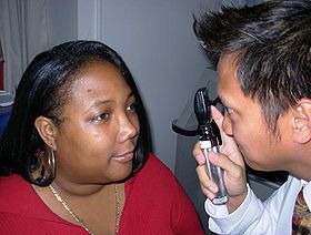
 Ophthalmoscopic exam: the medical provider would next move in and observe with the ophthalmoscope from a distance of one to several cm. | |
| MeSH | D009887 |
Ophthalmoscopy, also called funduscopy, is a test that allows a health professional to see inside the fundus of the eye and other structures using an ophthalmoscope (or funduscope). It is done as part of an eye examination and may be done as part of a routine physical examination. It is crucial in determining the health of the retina, optic disc, and vitreous humor.[citation needed]
The pupil is a hole through which the eye's interior can be viewed. For better viewing, the pupil can be opened wider (dilated; mydriasis) before ophthalmoscopy using medicated eye drops (dilated fundus examination). However, undilated examination is more convenient (albeit not as comprehensive), and is the most common type in primary care.
An alternative or complement to ophthalmoscopy is to perform a fundus photography, where the image can be analysed later by a professional.