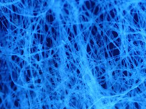
Autofluorescence is the natural emission of light by biological structures such as mitochondria and lysosomes when they have absorbed light, and is used to distinguish the light originating from artificially added fluorescent markers (fluorophores).[1]
The most commonly observed autofluorescencing molecules are NADPH and flavins; the extracellular matrix can also contribute to autofluorescence because of the intrinsic properties of collagen and elastin.[1]
Generally, proteins containing an increased amount of the amino acids tryptophan, tyrosine, and phenylalanine show some degree of autofluorescence.[2]
Autofluorescence also occurs in non-biological materials found in many papers and textiles. Autofluorescence from U.S. paper money has been demonstrated as a means for discerning counterfeit currency from authentic currency.[3]
- ^ a b Monici, M. (2005). "Cell and tissue autofluorescence research and diagnostic applications". Biotechnology Annual Review. 11: 227–256. doi:10.1016/S1387-2656(05)11007-2. ISBN 9780444519528. PMID 16216779.
- ^ Cite error: The named reference
Menter-2006was invoked but never defined (see the help page). - ^ Chia, Thomas; Levene, Michael (17 November 2009). "Detection of counterfeit U.S. paper money using intrinsic fluorescence lifetime". Optics Express. 17 (24): 22054–22061. Bibcode:2009OExpr..1722054C. doi:10.1364/OE.17.022054. PMID 19997451.
